This video will serve as an introduction to visual pattern recognition. Your chapter begins with a detailed description of the human visual system. And it uses agnosia to provide an example for how damage within different parts of the system can cause different perceptual problems. For example, patients with a perceptive agnosia load are believed to have problems with early processing of information in the visual system. These individuals will be unable to recognize very simple shapes. And then it also goes through associative agnosia, which in these individuals they have a hard time recognizing more complex objects. And they would be adoring, recognizes simple shape but not more complex objects. And that makes sense if you remember back to how the visual field, sorry, the visual information is processed in the brain. So in terms of the core texts, its first process, the occipital lobe. And from there, the same information takes two different streams. One, the dorsal stream and the second and the ventral stream. Now if you remember from physiological psychology, the dorsal stream, that information is more concerned with where an object is in space and how to manipulate it. And then eventual stream is more concerned with actually identifying an object or what is this object? So if a person has a perceptive agnosia, that the the damages probably the answer to your occipital lobe, right? So the damage is occurring at a very early local processing. However, if you have associative agnosia, you’re not you’re not able to identify complex objects. That damage is probably occurring somewhere in your ventral stream. And depending on what type of object should have the most trouble with, a lot of times they can pinpoint a specific area of this ventral stream. Ok, so just to review how information actually gets to the brain. This is a diagram of a human eye. And so light is going to ensure through the cornea. And it will, through the cornea, it’ll pass through the lens. And at this point, light is bent depending on the shape of the lens. And then it hits the retina. So if everything’s working the way it should, the lens should be focusing information onto the fovea. The fovea allows us to take full advantage higher at the high resolution power of the cones. Perceiving objects is information that is the focus on the foia is going to have the most sharpness. It’s also going to have the most color sensitivity as well. Whereas information follow falling on the peripheral part of the retina. This area here and down here is going to be less sharp. It’s going to be less sensitive to color. However, is going to be more sensitive to low light situations. So that this area where the fovea is located where you hadn’t most tones and your sharpest. As you can see, it’s actually very small compared to the rest of the retina. And if you take your arm and hand NGO, you hold an arm, arm’s-length out. You hold your fingers apart about the shape of a Great. So just pretend you’re holding agree between your thumb and your you’re pointing finger. That area is about the area that will fall on your retina. The rest of your vision is much less sharp. But because the, the brain is able to bind all that information together, you never realize that you, you think that everything is a sharp, is the information that falls on the fovea. So once visual information is transferred from lightwaves into neural impulses via the retina. There’s a signal from the retina that goes through the ganglion cells. And those ganglion cells, their axons that come your optic nerve. The optic nerve first passes through the thalamus and then makes its way to the cortex. Within the occipital cortex or your primary visual cortex. We have found there are receptive fields within that area. And receptive field just represents, is a representation of the information that it manipulating in the outside world. So when information falls on the retina, on the part of the retina is represented in the specific visual fields in the occipital cortex. Then these visual fields are activated and we have several different types of visual felt. So depending where the light falls in a cell’s receptive field, the cells, a rate of firing will increase, decrease, or stay the same. And these are known as on-off or off. On cells. Hubel and Wiesel discovered the visual cortex in cats. And they discovered that the view cortical cells have a long gated receptive fields that respond to more complex stimuli so that we have what we call edge detectors, for example. And these respond positively or negatively depending on where light falls. Relative to a line. So there’s just imagine a line down the middle of this receptive field. So flights falling on the right of that, it will either increase or decrease. And then if it’s falling on the left and that the same thing. While var detectors respond positively to, positively or negatively depending on where white falls in the center or on the periphery. Here you can see representation of those fields taken from your textbook. This is the edge detector field and you can see it can be, either of these can be activated by the left side or it can be inhibited by the left side by light falling on the left side. And then it’s going to be the opposite on the other side. And this is a representation of the bar receptive fields. So this is what I was saying earlier. You have this bar in the middle of the receptive field. And it’ll either be activated when light falls in this middle area or it’ll be inhibited and then the opposite on each side. So now to put this all together, you remember earlier we had gone off cells. So it’s either I turn on when light falls in the middle are off on the outer part or the opposite. We can put that together in these more complex receptive field. So it could be that these more complex receptive fields, the bar and the edge detectors, are actually made up of these on ourselves. And then finally, Hubel and Wiesel devise a hyper column representation as cells in the primary visual cortex with each heifer column comprised of cells that respond to different features of this receptive field. So you can see here these lines represent what this particular receptive field orientation of light that this particular receptive field is the most sensitive to and was truly amazing about this, is that it’s so well organized. So for every area in the retina, you have this representation of that area in the primary visual cortex. And then on top of that, you have these columns. So these columns are made up of cells. There’s layers of neurons here doing different things, but each of these columns made up of these layers of neurons are more sensitive to a specific orientation. And finally, all of these kind of micro organizational properties of the brain have been put together into these theories of perception. And the two main theories of perception discussed in this chapter are the template making and feature analysis. So with template matching. A retinal image of an object is faithfully transmitted to the brain. And the brain attempts to compare the image directly to various stored templates. So if you see a coffee cup, for example, you will compare that to all the other times you’ve seen a coffee cup and if it matches close enough, then you’re able to recognize it. The other is that feature analysis. And in this, stimuli are thought of as combinations or the elemental features. So it’s not that you’re looking at an object as a whole. It’s that you’re taking in a combination of features in trying to match that to a database that features that you’ve previously been exposed to. Okay, so one way to look at what can go wrong when these associative areas of the brain are lesion. This disorder called prosopagnosia. And it comes from it comes from a lesion either due to an accident or a stroke, or some individuals are born with us. But illusion to the fusiform gyrus. So just to orient you to this picture, this is a whole brain. Split it down the middle UV looking at the inside of the brain, right? So what we call a midsagittal view, that the brain, if you flip it over so that you’re looking at the bottom of the brain. That’s, This is what you would see the fusiform gyrus in the temporal lobe of the brain. When you have a lesion of this area and people have a really hard time recognizing faces. Now when I see that you might think that it looks something like this. And that’s not really the case because here, you can’t see any of the features. If you, if a person has prosopagnosia and you say OK, how many ices first an app does this person never knows him out? They can racking gnosis features. They see those features, but they can put them together in a meaningful way. So that’s, there’s a higher area of processing which the fusiform gyrus, which is what we think the fusiform gyrus is doing. Okay, so just to give you an idea, let’s look at three pictures and some celebrities. Now you may recognize one or a couple of these and they take you a few seconds, Take your time trying to figure out who they are. Alright, now look at the same people. So you probably recognize all of these guys instantly, right? This is Prince Henry, Sandra Bullock, and Bill Murray. So why was it so hard when they were upside down? And the reason is because your fusiform gyrus finely tuned to the way that human faces are normally oriented. So when you saw it like this, you could still see their eyes, you see their nose or mouth. You could see all the features. But it didn’t have meaning the way it does when you see it this way. Now, other objects don’t work that way. You take a pen or pencil turn upside down as still pen or pencil, you recognize it and still a, you can take a coffee mug and do the same thing, nearly anything except for human faces. And that’s because the fusiform gyrus is finely tuned to recognizing faces and their normal orientation. I put you through this exercise though, so that you would have an idea of what prosopagnosia is like. You can see all the features that they don’t have any meaning. And it says higher cognitive processing that’s responsible for giving it meaning and helping you recognize this objects.
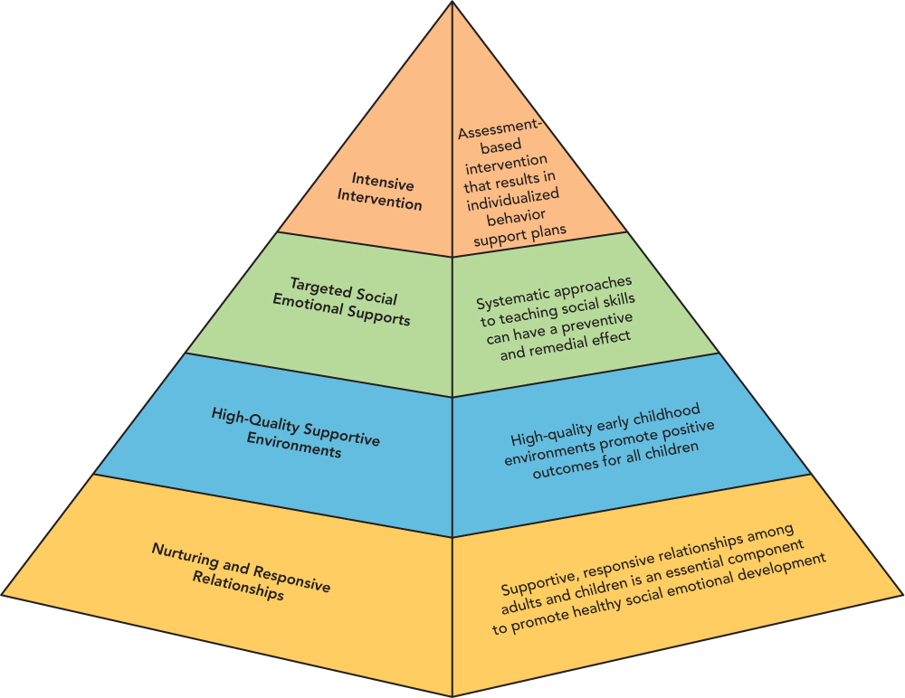
 Source: From Promoting Social and Emotional Competence in Infants and Young Children. The Center on the Social and Emotional Foundations for Early Learning, http://csefel.vanderbilt.edu. Reprinted by permission.